Radiographer role: responsibilities, skills and workplace safety
A radiographer does much more than operate an X-ray machine. They help clinicians see what is happening inside the body, support patients through unfamiliar procedures and make sure imaging is carried out safely. It is a role where technical precision and human kindness need to work side by side: the image must be clear, but the person behind it matters just as much.
For healthcare employers and safety professionals, understanding the radiographer’s responsibilities is essential to creating a safe, compliant imaging service. Here is what the role involves, which skills matter most and how radiographers help manage workplace risks every day.
What does a radiographer do?
Radiographers are registered healthcare professionals who use imaging equipment to produce diagnostic images or deliver certain treatments. Diagnostic radiographers commonly work with X-rays, computed tomography (CT), magnetic resonance imaging (MRI), ultrasound and fluoroscopy. Therapeutic radiographers use radiation to treat conditions such as cancer.
The exact duties vary by specialism and workplace. A radiographer in a hospital emergency department may need to image a suspected fracture quickly, while someone working in a cancer centre may help plan and deliver a carefully controlled course of radiotherapy. In both settings, the aim is to provide high-quality care while managing clinical and workplace risks.
Radiographers work closely with patients, radiologists, doctors, nurses, medical physicists and other professionals. They may also help explain procedures and prepare patients, but they do not replace the clinician who interprets findings and makes a diagnosis.
Core responsibilities in diagnostic imaging
Before an examination, the radiographer checks the request and confirms that the proposed imaging is appropriate for the patient and clinical question. They identify the patient using suitable checks, explain what will happen and consider relevant information such as mobility needs, previous reactions to contrast media or the possibility of pregnancy.
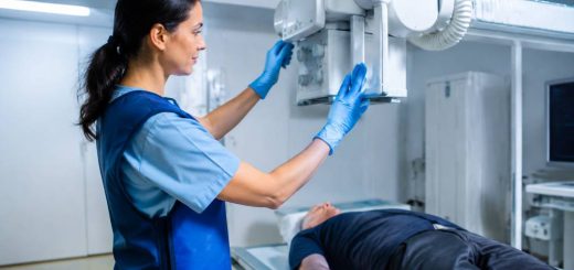
During the examination, they position the patient and equipment, select suitable imaging settings and give clear instructions. A small adjustment to positioning can make the difference between a useful image and a repeat scan. And repeat exposures are not anyone’s favourite way to spend an afternoon.
After the images are acquired, the radiographer checks them for quality and confirms that the required views have been obtained. Depending on local procedures and their training, they may carry out additional checks, flag urgent concerns through agreed pathways or provide further information to the reporting clinician.
Other responsibilities can include:
- Maintaining accurate patient and examination records.
- Preparing and checking imaging rooms and equipment.
- Following infection prevention and control procedures.
- Supporting patients who are distressed, in pain or unable to move easily.
- Reporting equipment faults, near misses and safety concerns.
- Helping maintain a respectful, private and accessible environment.
Radiation safety: careful use, not guesswork
Ionising radiation is valuable in diagnosis and treatment, but it needs to be managed properly. In the UK, work involving medical exposures is governed by regulations including the Ionising Radiation (Medical Exposure) Regulations 2017, commonly known as IR(ME)R. Workplace protection from ionising radiation is also addressed by the Ionising Radiations Regulations 2017 (IRR17).
These regulations place duties on employers and other defined duty holders. The precise responsibilities depend on the setting and the individual’s role, so radiographers should follow their employer’s procedures and training rather than rely on assumptions. In practice, this means checking that the exposure is appropriately authorised, using the correct protocol and taking steps to keep exposure as low as reasonably practicable while still achieving the clinical purpose.
Radiographers use measures such as appropriate collimation, suitable exposure settings, correct positioning and room shielding. They also make sure people who do not need to be in the controlled area are kept out during an exposure. Where staff must be present, local procedures, risk assessments and protective measures apply.
Consider a child who needs an X-ray after a fall. The radiographer may adapt the technique and positioning for the child’s size, explain the process in reassuring language and check that the image includes only the area needed. That combination supports a useful image while avoiding unnecessary exposure. Safety is not a separate step at the end; it is built into the examination.
Different equipment, different hazards
Radiation safety is only one part of the picture. Each imaging environment comes with its own risks, and a safe radiographer stays alert to the hazards that are easy to overlook when a department is busy.
- MRI: The powerful magnetic field can attract ferromagnetic objects and create serious hazards. Screening patients, staff and visitors is essential. Equipment and accessories must be suitable for the MRI environment, and access controls must be followed.
- CT and fluoroscopy: These use ionising radiation. Staff need to follow local protocols, use the room correctly and apply relevant protective measures, particularly when working with fluoroscopy.
- Ultrasound: Although it does not use ionising radiation, prolonged scanning and awkward posture can contribute to musculoskeletal problems. Good workstation setup and sensible breaks matter.
- Contrast procedures: Where contrast media are used, staff follow local checks and emergency procedures, including steps to respond to adverse reactions.
One familiar near miss in MRI departments is a visitor entering with an everyday metal item they had no reason to think about: a tool, a set of keys or a portable oxygen cylinder. Thorough screening and controlled access may feel repetitive, but repetition is precisely what helps prevent an avoidable incident.
Patient handling, infection control and everyday safety
Many patients arrive in pain, with limited mobility or attached to monitoring equipment. Helping someone onto an imaging table can therefore involve risks to both the patient and staff. Radiographers should assess what assistance is needed, use suitable transfer aids and ask colleagues for help when required. Trying to “just manage” a difficult transfer is not a badge of honour; it is a preventable injury waiting for a convenient moment.
Good infection control is equally practical. Reusable equipment and contact surfaces need to be cleaned in line with local procedures, especially when patients move between clinical areas. Hand hygiene, appropriate personal protective equipment and safe handling of contaminated items help protect patients and staff alike.
Other routine workplace risks include slips and trips from cables, cluttered rooms, electrical equipment faults and lone working in smaller or out-of-hours services. Clear walkways, regular equipment checks and a straightforward method for raising concerns make a real difference. A hazard reported early is much easier to manage than one discovered by someone’s ankle.
Skills that make a difference
Technical knowledge is essential, but it is only one part of effective practice. Radiographers need to combine clinical judgement with communication, attention to detail and a calm approach under pressure.
- Communication: Patients may be anxious, confused or in pain. Clear explanations and a chance to ask questions can make an unfamiliar procedure feel more manageable.
- Attention to detail: Confirming identity, checking the requested examination and selecting the right protocol all support safe care.
- Adaptability: A patient may be unable to hold a position or follow a standard instruction. Radiographers need to adjust their approach without losing sight of the clinical need.
- Teamwork: Imaging often depends on coordinated work between clinical teams, support staff and technical specialists.
- Risk awareness: Recognising a potential issue, pausing when necessary and escalating concerns are important professional skills.
- Emotional resilience: Radiographers may work with seriously ill patients and distressed families. Compassionate care needs support, reflection and healthy workplace practices.
These skills often come together in a simple moment. If a patient is too frightened to lie still for a scan, the best response may not be to repeat instructions more loudly. A calm explanation, a small adjustment to positioning or a brief pause may improve both the patient’s experience and the quality of the images.
Training, registration and professional development
In the UK, radiographers complete an approved training route and must be registered with the Health and Care Professions Council (HCPC) to practise under the protected title. They are also expected to keep their knowledge and skills up to date through continuing professional development and to work within their competence.
For employers, this means supporting more than initial induction. Staff need role-specific training on equipment, local procedures, emergency arrangements and relevant safety requirements. Changes to software, protocols or equipment should be accompanied by appropriate instruction, not a hurried tour and a hopeful “you’ll pick it up”.
Supervision and clear escalation routes are particularly important for students and newly qualified staff. They should know who to contact if a request is unclear, equipment behaves unexpectedly or a patient’s condition changes during an examination.
How employers can support safer radiography
Radiographers carry out many safety checks, but they cannot create a safe service on their own. Employers and managers have a key role in providing suitable equipment, staffing, training and systems for reporting and learning from incidents.
Practical steps include:
- Keeping risk assessments and local procedures current and accessible.
- Maintaining equipment and acting promptly on reported faults.
- Providing appropriate training for radiation, MRI, manual handling and infection control risks.
- Making incident and near-miss reporting straightforward and encouraging a learning-focused response.
- Reviewing workloads, room layouts and staffing to identify avoidable pressure points.
- Supporting staff wellbeing, including access to supervision and opportunities to discuss challenging cases.
For example, if staff repeatedly report that a CT room becomes congested during patient transfers, the answer may involve reviewing the layout, storage or handover process—not simply reminding everyone to be more careful. Reliable safety improvements often come from fixing the conditions that make errors more likely.
Putting safe, compassionate imaging into practice
A radiographer’s work brings together technical skill, patient care and risk management. From confirming a patient’s identity to checking MRI access, positioning someone with limited mobility or reporting a faulty piece of equipment, each decision contributes to a safer service.
For healthcare teams, the most useful question is not whether every risk can be removed. It is whether the right people, procedures and support are in place to identify risks early and manage them consistently. When they are, radiographers can focus on what they do best: producing useful images and helping patients feel cared for throughout the process.