


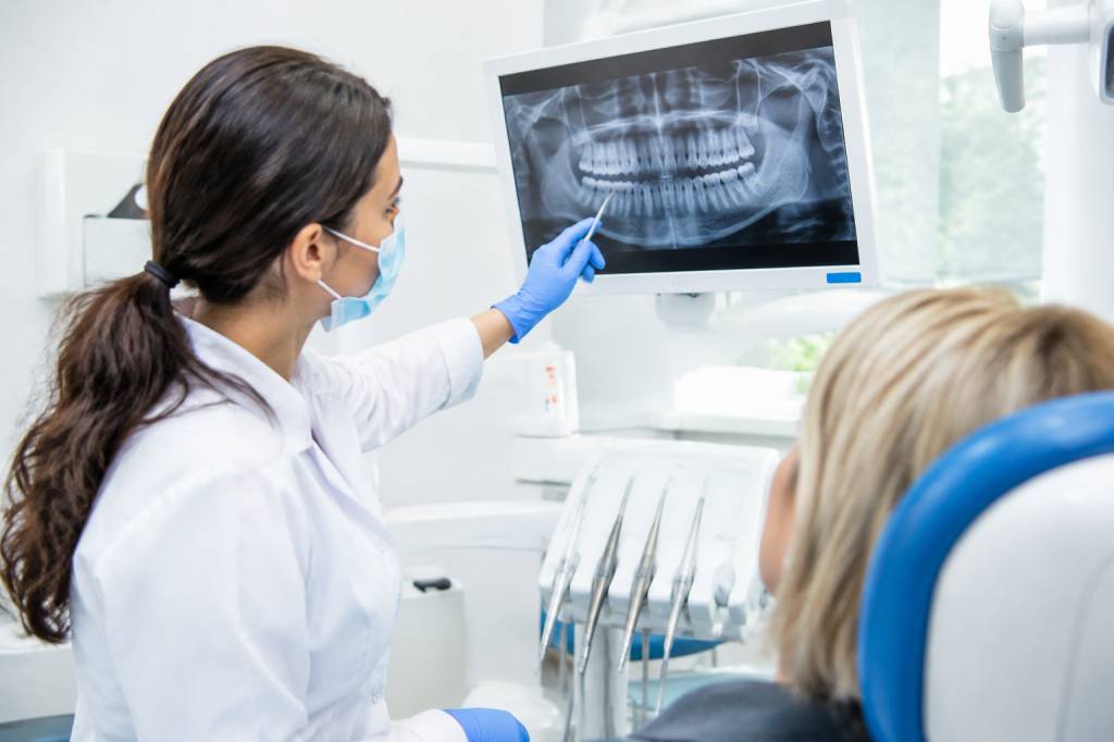
Dental imaging helps clinicians diagnose problems that are difficult to spot during a visual examination. But every X-ray also involves ionising radiation, equipment, infection-control considerations and a busy team working around patients. Safe imaging is not simply a matter of pressing the exposure button and hoping for the best.
For dental practices, good workplace procedures protect patients and staff while helping images answer the clinical question first time. This guide covers the practical steps: choosing the right examination, preparing the room, positioning safely and learning from near misses. Because a clear image is useful; a clear image taken safely is better.
Start with a clinical reason
The first safety check happens before anyone enters the imaging room: is an image needed, and will it help with this patient’s care? Dental radiographs should be requested on an individual basis, following an appropriate clinical assessment and the relevant professional guidance. A routine image “because it is time” may not be justified if it will not influence diagnosis or treatment.
Under the UK’s Ionising Radiation (Medical Exposure) Regulations 2017, commonly known as IR(ME)R, medical exposures must be justified and optimised. In practice, the people involved have defined responsibilities. The referrer requests an examination, the practitioner justifies it, and the operator carries out the practical aspects. One person may hold more than one role, depending on their training and local arrangements, but the responsibilities still need to be understood.
Before imaging, check the patient’s history and the clinical question. Is the aim to investigate pain, assess a tooth before treatment, or review a specific finding? Could previous images answer the question? Avoid repeating an image simply because it is convenient. A quick check of the records can save the patient an unnecessary exposure and the team a repeat appointment.
Know your legal and local responsibilities
Dental employers need suitable arrangements for radiation protection, including risk assessment, written procedures and staff training. The detail depends on the equipment and how the practice operates. Practices should follow current legislation and guidance, and seek advice from their Radiation Protection Adviser (RPA) and, where required, Medical Physics Expert (MPE).
Keep the practice’s written procedures and local rules accessible to the people who need them. These should explain how imaging is requested and authorised, who may operate each unit, what checks are required, and what to do if something goes wrong. Staff should know where the documents are—not just that they exist somewhere in a folder with a label last updated several years ago.
Local rules should reflect the actual room, equipment and working arrangements. They may identify controlled areas, outline access restrictions during exposure and specify who is responsible for maintaining radiation protection measures. Do not assume that a procedure written for one room or machine automatically suits another.
Prepare the room and equipment
Before the patient arrives, check that the equipment is ready and the room can be used safely. Make sure the imaging unit appears undamaged, the positioning aids are available, and the exposure controls work as expected. Follow the manufacturer’s instructions and the practice’s quality assurance schedule. If a unit is faulty, behaving unusually or producing inconsistent images, take it out of use where appropriate and report it through the practice’s maintenance process.
Plan the exposure so staff can operate the equipment from the designated protected position, in line with local rules. Keep unnecessary people out of the room during exposure and make sure the patient can hear and follow instructions. Before pressing the control, check that the patient is correctly positioned, the selected settings and examination are appropriate, and no one is in an unsafe location.
Good housekeeping matters too. Cables, bags and equipment left in walkways can turn a straightforward appointment into a trip hazard. Keep the room tidy, but do not move protective barriers or alter the layout in ways that conflict with the radiation risk assessment.
Position for a useful image first time
Many repeat exposures happen because of positioning errors, movement or incorrect selection—not because the machine has developed a mysterious dislike of Tuesdays. A calm explanation and a few seconds of preparation can make a real difference.
Explain what the patient needs to do in plain language. For an intraoral image, tell them where the receptor will sit and ask them to remain still. For panoramic imaging, explain how to stand, where to place their chin and hands, and why they may need to remove objects such as earrings or glasses. Use the equipment’s positioning guides and follow local procedures rather than relying on memory alone.
Check that the examination selected on the console matches the request. Make sure the patient’s position is comfortable enough to maintain, and consider whether they may need additional support or adapted instructions. Children, people with mobility limitations and patients experiencing anxiety may need more time or a different approach. Never rush a patient into a position they cannot safely hold.
If an image is not diagnostically acceptable, assess why before repeating it. A repeat should be based on clinical need and local procedures, not an automatic response to every imperfection. Recording and reviewing repeat-image causes can reveal patterns: perhaps one positioning aid is difficult to use, a particular setting is being misunderstood, or staff would benefit from refresher training.
Protect staff without compromising care
Staff should not hold a patient or receptor during an exposure unless there is an exceptional, justified reason and local procedures permit it. First consider whether the patient can be supported using a positioning aid or another safe method. If assistance is genuinely necessary, follow the practice’s radiation protection procedures and ensure the person helping is appropriately instructed and protected.
Do not use a member of staff as a routine substitute for a suitable positioning aid. A colleague’s willingness to help does not remove the employer’s duty to control exposure. Occupational monitoring arrangements, including personal dosimetry where indicated by the risk assessment or advice from the RPA, should be followed. Staff should wear and store dosimeters as instructed; a badge left in a drawer is not much of a monitoring programme.
Pregnancy should be handled sensitively and in accordance with the employer’s radiation protection arrangements and applicable guidance. Staff should know how to raise a concern confidentially and what individual risk assessment or work adjustments may be appropriate. Avoid blanket assumptions: decisions should be based on the actual work, exposure risks and competent advice.
Build infection control into imaging
Imaging equipment is part of the clinical environment, so infection prevention cannot be an afterthought. Clean and disinfect contact surfaces between patients according to the manufacturer’s instructions and the practice’s infection-control procedures. Use appropriate barriers where suitable, and change them between patients. A barrier is not a substitute for cleaning when the equipment or guidance requires it.
Reusable positioning devices and receptors should be reprocessed using the correct method for that item. Follow the manufacturer’s instructions, including any limits on immersion or use of particular disinfectants. Keep clean and contaminated items separate, and make sure staff know where used items should go.
Hand hygiene and appropriate personal protective equipment remain essential. The precise PPE depends on the task and the practice’s infection-risk assessment. These small, repeatable steps protect patients and staff—and are far easier than trying to remember who touched which surface after a busy morning clinic.
Choose the right technology for the question
Dental practices may use intraoral, panoramic and cone beam computed tomography (CBCT) imaging. Each has a role, but they are not interchangeable. The right choice depends on the clinical question and the information needed. CBCT can provide detailed three-dimensional views, but its use should be justified for the individual patient and the smallest appropriate field of view selected in accordance with professional guidance and local protocols.
Staff need training and authorisation appropriate to the equipment they use. A familiar control panel does not automatically mean someone is competent to operate every imaging modality. Practices should also make sure images are assessed by an appropriately qualified practitioner and that findings are communicated and followed up through a reliable process.
Digital systems bring practical benefits, including fast image access, but they also require sensible information handling. Store images in the approved clinical record system, restrict access appropriately and follow the practice’s data-protection and retention policies. Confirm the correct patient record before saving an image; a technically perfect radiograph in the wrong file creates a very different kind of problem.
Make quality assurance part of the routine
Quality assurance is not a once-a-year box-ticking exercise. A planned programme helps confirm that equipment and processes continue to perform as expected. It may include routine visual checks, image-quality review, monitoring of repeat rates, equipment maintenance and periodic testing in line with current requirements and advice from competent specialists.
Assign clear responsibility for checks and record the results. If a check identifies a problem, document what was found, who was informed and what action followed. Reviewing repeat images is particularly useful when it is done constructively. The aim is to improve the system, not to shame the person who had a difficult appointment with a patient who could not keep still.
Use findings to update training and procedures. For example, if a review shows repeated positioning errors with one type of panoramic examination, arrange a short practical refresher and check whether the instructions or equipment setup can be improved. Small adjustments can prevent a long chain of repeat exposures.
Respond well when something goes wrong
Incidents and near misses should be reported through the practice’s established process. Examples might include an unintended exposure, equipment malfunction, an exposure involving the wrong patient, or a failure to follow a radiation protection procedure. Staff should know whom to contact and what information to record.
Do not try to quietly fix an incident by repeating an exposure or deleting a record. Follow the practice’s incident procedure, involve the appropriate radiation protection contacts and assess whether the event meets the criteria for escalation or reporting under relevant regulations. Prompt, accurate documentation helps the practice respond properly and identify what needs to change.
A blameless learning culture is important. If people fear embarrassment, they may keep quiet about near misses—exactly the events that can reveal a weakness before someone is harmed. Ask practical questions: Was the procedure clear? Was the equipment working? Did workload or room layout contribute? What would make the safe action easier next time?
Make safer imaging everyone’s business
Effective dental imaging depends on a chain of decisions: justifying the examination, preparing equipment, positioning the patient, controlling exposure, maintaining infection control and reviewing image quality. If one link is weak, both safety and clinical value can suffer.
Start with a simple team check-in. Do staff know their IR(ME)R responsibilities? Are local rules easy to find? Can everyone explain how to report an equipment concern or near miss? A short conversation can uncover gaps before the next patient arrives.
When procedures are practical, training is current and staff feel able to raise concerns, safer imaging becomes part of everyday care—not an extra task squeezed between appointments. That is good for patients, good for the team and, importantly, good for getting the image right first time.