


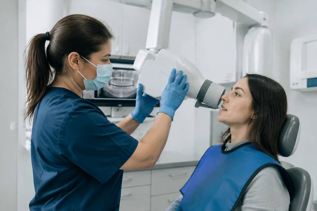
Dental radiography may take only a few minutes, but getting a useful image safely depends on much more than pressing a button. The person taking the X-ray needs appropriate training, a clear understanding of their responsibilities and a reliable approach to radiation protection.
In the UK, “dental radiographer” is not usually a separate protected job title. Dental nurses, dentists, dental hygienists and dental therapists may take dental radiographs when they are properly trained, entitled to act as an operator and working within their professional scope. Here is what that means in practice, from training and daily checks to patient care and compliance.
What does a dental radiographer do?
A dental radiographer operates dental X-ray equipment to produce images that help a suitably qualified dental professional assess a patient’s oral health. These might be bitewing images for checking between teeth, periapical images showing a tooth and its root, or panoramic images that capture a wider view of the jaws.
The role is not simply technical. A good operator helps the patient understand what will happen, positions them carefully, selects the correct equipment settings and checks that the resulting image is useful. They also follow local radiation protection procedures and help keep accurate records.
Imagine a patient who is nervous about an intraoral X-ray and keeps moving just as the exposure is about to happen. Clear, calm instructions and a little reassurance can make the difference between a diagnostic image and a repeat exposure. In radiography, communication is part of safety—not an optional extra.
Who can take dental X-rays in the UK?
Several dental professionals may take radiographs, but they must meet the requirements of the Ionising Radiation (Medical Exposure) Regulations 2017, commonly known as IR(ME)R. Under these regulations, people involved in a medical exposure have defined responsibilities. The main roles include:
- Employer: The organisation responsible for putting the required procedures, training and safety arrangements in place.
- Referrer: A professional who requests an exposure and provides relevant information to support the decision.
- Practitioner: A suitably entitled professional responsible for justifying the exposure and taking responsibility for the clinical decision.
- Operator: The person who carries out practical aspects of the exposure, such as positioning the patient and operating the equipment.
One person may perform more than one role if they are appropriately trained and entitled to do so. The important point is that responsibilities must be clear. Being confident with the equipment does not, by itself, make someone entitled to carry out every part of the radiographic process.
Dental professionals should also work within their registration requirements, professional guidance and the procedures set by their employer. If you are unsure whether a task falls within your role, check your training record, local rules and the advice of your practice’s radiation protection lead before proceeding.
Training and qualifications
Anyone taking dental radiographs needs education and training that matches the type of equipment and exposures they will carry out. A dental nurse, for example, generally needs appropriate theoretical and practical training in dental radiography before taking images as an operator. Recognised routes may include a post-registration dental radiography qualification or a course accepted by the relevant employer and professional framework.
Training should cover more than button-pressing. It commonly includes:
- The principles of radiation protection and the risks associated with ionising radiation.
- Relevant legislation, including IR(ME)R, and the responsibilities of each role.
- How to select the correct image type and equipment settings under local procedures.
- Patient identification, communication, positioning and assistance.
- Image quality, common positioning errors and when an image needs further review.
- Equipment checks, fault reporting, record keeping and local radiation protection rules.
Practical competence matters as much as classroom knowledge. A trainee should be supervised until they can demonstrate that they can perform the task safely and consistently. The employer must ensure that operators are adequately trained and entitled for the work they undertake; a certificate is valuable evidence, but it does not replace workplace procedures or continuing competence.
Training is not a one-and-done event. Equipment, guidance and clinical protocols can change, and skills can become rusty. Regular updates, reflection on image quality and appropriate continuing professional development help keep practice current. If a colleague has not taken a particular type of radiograph for some time, a refresher or supervised return to practice may be sensible.
Safety before, during and after an exposure
The central aim is to obtain the information needed for patient care while keeping exposure as low as reasonably practicable. This starts before the patient enters the imaging room. The referrer and practitioner must follow the appropriate process for requesting and justifying an exposure, and the operator must check that the request and patient details are correct.
Before taking an image, confirm the patient’s identity, explain the procedure and check that they understand what is required. Use the correct imaging technique and settings for the patient and clinical need, following local protocols. Do not take an exposure simply because it is convenient or because a routine appointment usually includes one: there should be a clear clinical reason.
During the exposure, position the patient and receptor carefully, use the equipment as trained and follow the practice’s rules for staff positioning. The operator should not hold the receptor or X-ray tube head during an exposure. Staff should use the designated protective area or stand at the specified distance and angle in accordance with local rules.
Protective equipment requirements can vary by situation and current guidance. Do not rely on habit or assumptions about lead aprons and thyroid collars: follow your employer’s procedures and current professional advice. Good technique and avoiding unnecessary exposures are fundamental controls, not substitutes for protective equipment where it is required.
After the exposure, check that the image is of diagnostic quality and ensure it is available for the appropriate clinical review. If an image is not satisfactory, consider why before repeating it. Was the receptor misplaced? Did the patient move? Could clearer instructions or a different technique solve the problem? Repeats should be justified, not automatic.
Patient care and communication
Patients may be worried about radiation, discomfort or the possibility of a gag reflex. A brief explanation can prevent a lot of uncertainty: tell them what the receptor will feel like, how long they need to stay still and how they can signal if they need a pause. Small adjustments, such as giving the patient time to practise biting gently on the holder, can make the experience easier.
Check whether the patient needs additional support. Children, people with learning disabilities, patients with limited mobility and those who speak little English may need a different approach. Use suitable communication aids or an interpreter where appropriate, and follow safeguarding and consent procedures. Never rush a patient into an exposure they do not understand.
Questions about pregnancy should be managed according to current guidance and local procedures. Pregnancy is not automatically a reason to withhold a clinically justified dental radiograph, but the exposure must still be justified and optimised. If there is uncertainty, pause and seek advice from the practitioner or the person responsible for radiation protection.
Equipment, quality assurance and records
Safe radiography depends on reliable equipment and a working quality assurance programme. Practices should carry out the checks and testing required by their procedures, keep maintenance and test records, and make sure faults are reported promptly. If equipment appears damaged or is producing inconsistent images, do not quietly work around the problem; follow the practice’s fault-reporting process.
Good records support both patient care and compliance. Depending on local procedures, these may include the clinical reason for the exposure, the image taken, relevant operator details, quality assurance information and any repeat exposure. Documentation should be accurate and completed at the time, rather than reconstructed later from memory.
Local rules should be accessible to staff who work with the equipment. They explain how radiation protection is managed in that particular setting, including designated areas, safe working arrangements and what to do if something goes wrong. Staff should know where to find them and whom to contact with questions.
Common mistakes—and how to avoid them
Many problems are preventable and do not require complicated solutions. A few habits make a substantial difference:
- Taking an image without a clear clinical reason: Confirm that the exposure has been appropriately requested and justified.
- Using the wrong settings or technique: Follow the protocol for the equipment and patient, and seek help if the situation falls outside your training.
- Repeating an image without reviewing the cause: Identify the error first and adjust the approach before any repeat.
- Assuming experience equals entitlement: Check that your training and authorisation cover the task you are performing.
- Skipping checks when the practice is busy: Build equipment and patient checks into the routine. A hectic diary is not a radiation protection strategy.
- Leaving procedures unread or outdated: Review local rules and updates when equipment or practice arrangements change.
For example, if bitewing images repeatedly show overlapping contacts, the answer may not be “try again and hope”. Reviewing horizontal angulation, receptor placement and patient instructions can identify the cause and improve the next attempt. That saves time and avoids unnecessary exposure—a win for the patient and the diary.
Building a strong safety culture
Radiation protection works best when it is treated as a shared responsibility. Employers need suitable procedures, equipment and training; practitioners need to justify exposures appropriately; operators need to work within their competence and follow local rules. Everyone should feel able to raise a concern without worrying that they are being difficult.
Dental teams can strengthen practice by reviewing image quality, discussing repeat rates and using incidents or near misses as learning opportunities. A useful question after a difficult exposure is not “Who got it wrong?” but “What would make the safe approach easier next time?” That shift encourages honest reporting and practical improvements.
For anyone considering this career path, dental radiography offers a valuable blend of patient interaction, technical skill and responsibility. The best operators combine careful positioning with clear communication and sound judgement. When training, safety procedures and teamwork are in place, a short exposure can provide useful clinical information with confidence and care.