


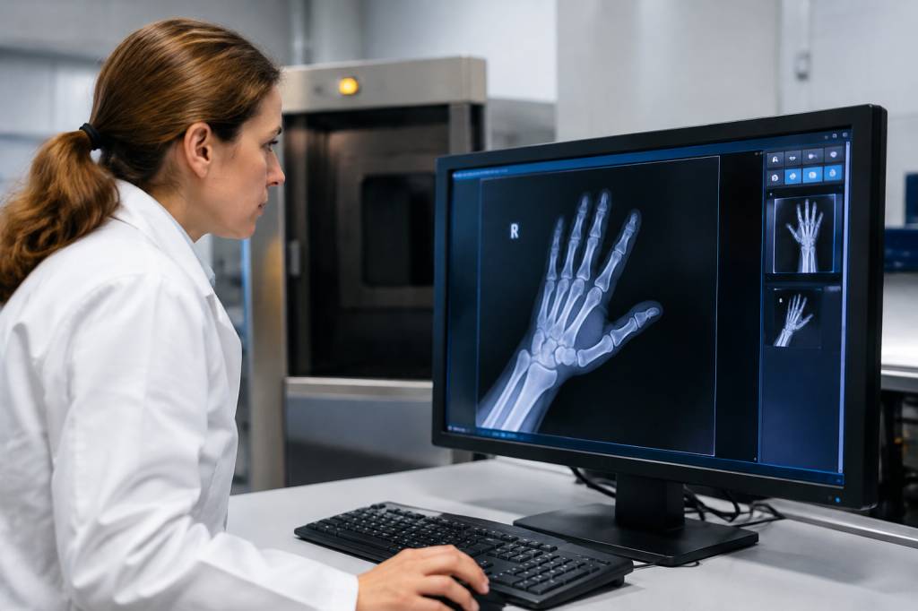
When a workplace incident leaves questions unanswered, the most important evidence is not always visible to the naked eye. A damaged machine may conceal a fractured component. An injury may involve an object or pattern that is difficult to assess from an external examination alone. In these cases, forensic radiography can help investigators look beneath the surface—without disturbing the evidence first.
It is a specialist field, most often associated with forensic medicine and criminal investigations. But its methods can also inform health and safety inquiries, particularly when understanding an injury, a failed piece of equipment or the sequence of events matters. Used appropriately, radiography can add detail to an investigation. Used carelessly, it can create false confidence or compromise evidence. The difference lies in planning, expertise and a clear chain of custody.
What is forensic radiography?
Forensic radiography uses imaging techniques to document and examine evidence for investigative purposes. The familiar example is an X-ray, but the field can also include computed tomography (CT), digital radiography and, where appropriate, other imaging methods.
In healthcare, imaging is generally performed to help diagnose or treat a patient. In a forensic setting, the priorities are different: preserve the original state of the evidence, create an accurate record, answer a defined investigative question and ensure that images can be trusted if they are later reviewed or presented in formal proceedings.
That evidence might be a human injury, a safety helmet, a section of machinery, a tool or an object recovered from an incident scene. The radiographer’s role is not to decide who is at fault. It is to produce images that may help qualified investigators, clinicians or forensic specialists understand what is present.
Where it can support a health and safety investigation
Forensic imaging is not necessary for every workplace incident. A minor trip, for example, is unlikely to call for a CT scanner. But where evidence is complex, hidden or easily damaged, imaging may help answer questions that photographs and visual inspection cannot.
-
Injury assessment: Imaging may help document fractures, foreign objects or other features relevant to understanding how an injury occurred. Clinical care must always come first; a forensic purpose should never delay necessary treatment.
-
Equipment failure: X-ray imaging can reveal internal cracks, voids, weld defects or damage within certain components without cutting them open. This may help preserve an item for further examination.
-
Protective equipment: A helmet, glove or other item may be imaged to document internal damage, embedded fragments or deformation. The findings should be considered alongside inspection, testing and the user’s account.
-
Incident reconstruction: Images may help clarify the position of objects or the presence of concealed materials. They are one source of evidence, not a complete account of what happened.
-
Documentation: A high-quality image can provide a lasting record of an item’s condition before it is opened, dismantled or otherwise altered.
Imagine a worker is injured when a powered tool breaks during use. A visual check reveals a damaged housing, but the cause remains uncertain. Radiography might show an internal fracture or a manufacturing defect. That could help direct a more detailed engineering examination. It would not, on its own, establish whether the tool was poorly maintained, misused or defective from the outset. That requires context, records and expert interpretation.
Common techniques and what they can show
Conventional X-ray imaging passes a controlled amount of radiation through an object to create an image based on how different materials absorb it. Dense materials generally appear differently from less dense ones. This can make X-rays useful for locating metal fragments, examining some fractures and assessing the internal structure of suitable objects.
Digital radiography captures images electronically rather than on traditional film. Digital systems can make it easier to store, share and review images, but they do not remove the need for careful positioning, appropriate exposure and secure file handling. A sharp-looking image is not automatically a useful one.
Computed tomography takes multiple X-ray measurements from different angles and combines them into cross-sectional images, which can also be used to create a three-dimensional view. CT may be valuable where overlapping structures make a standard X-ray difficult to interpret or where an internal feature needs to be localised. It is usually more resource-intensive, so the investigative question should justify its use.
Specialist industrial radiography can be used to examine certain components and materials for internal defects. The method and equipment must be chosen by competent professionals, with suitable controls for the material, shape and size of the item. Not every object can be reliably assessed with the same technique.
Imaging has limits. Materials can overlap on a two-dimensional image; some defects may be too small or poorly positioned to detect; and an image can show that a feature exists without explaining how or when it formed. Radiography is a window into evidence, not a time machine.
Getting the process right
A useful examination starts with a specific question. “Can you X-ray this?” is less helpful than “Can imaging determine whether this component contains an internal fracture near the point of failure?” A focused question helps the specialist select the method, positioning and settings most likely to produce meaningful results.
Before imaging begins, investigators should record the item’s condition, identify it clearly and consider whether handling or transport could alter it. If the object is part of an active investigation, the relevant evidence-handling procedures should be followed from the outset.
-
Define the question the imaging is intended to answer.
-
Use a suitably qualified radiographer or other imaging specialist with experience relevant to the evidence.
-
Photograph and document the item before examination, where appropriate.
-
Record who handled the item, when it was transferred and how it was stored.
-
Retain original image files and record relevant examination details, including equipment and settings where applicable.
-
Document any processing or enhancement, and preserve the unaltered originals.
-
Make sure findings are interpreted by someone with the right expertise for the question being asked.
Chain of custody is not glamorous, but it is the investigative equivalent of labelling your lunch in a shared fridge: it helps everyone know what they are looking at and where it has been. More seriously, a clear record supports confidence that the evidence has not been substituted, lost or changed without documentation.
Radiation safety and ethical considerations
Radiography involves ionising radiation, so examinations must be planned and controlled by competent people. The type of equipment and setting will determine the relevant safety arrangements. These may include restricting access, controlling the exposure area, using suitable shielding and following established procedures. Workplace teams should never attempt to operate radiographic equipment without the required training, authorisation and safeguards.
When imaging involves a person, dignity, consent and clinical responsibility matter. The individual’s immediate health needs take priority, and examinations should be carried out by appropriate professionals in a suitable setting. Investigators should also consider privacy: medical images are sensitive information and should only be accessed, stored and shared for legitimate, authorised purposes.
For objects and equipment, ethical practice still matters. An examination should be proportionate to the question and should avoid unnecessary damage or exposure. If an item may be needed for further testing, investigators should agree in advance how imaging will fit into the wider examination plan.
Interpreting images without jumping to conclusions
One of the biggest risks in any investigation is confirmation bias: once a theory takes hold, people may start seeing every new detail as proof. Radiographic images are no exception. A mark, crack or fragment may look significant, but its meaning depends on the item’s history, the imaging method and other evidence.
For example, an internal crack in a machine part might have developed before the incident, during the failure or as a result of the impact afterwards. Imaging may locate the crack, but an engineering assessment and a review of maintenance records, operating conditions and witness accounts may be needed to interpret it.
Investigators should distinguish clearly between what an image shows and what they infer from it. “A fracture is visible in this area” is an observation. “The part failed because maintenance was inadequate” is a wider conclusion that requires supporting evidence. Keeping those statements separate makes reports more balanced and more useful.
Bringing imaging into a broader safety review
Forensic radiography can help establish facts, but preventing a repeat incident requires looking beyond the image. A complete health and safety review may also consider risk assessments, training, supervision, equipment inspection, work procedures, environmental conditions and worker feedback.
Suppose imaging finds a defect inside a lifting component after a near miss. The immediate response may involve securing the item and checking similar equipment. The longer-term review should ask how the defect developed, whether inspection arrangements could have detected it and whether the task exposed people to unnecessary risk. The aim is not simply to identify a damaged part; it is to understand how the safety system can be strengthened.
That is where forensic imaging earns its place: as one carefully chosen tool within a wider evidence-led investigation. It can reveal what was hidden, preserve a record before further examination and help experts ask better questions. The strongest investigations combine those images with sound evidence handling, competent interpretation and a genuine commitment to learning.