


Why imaging matters in occupational health
Imaging can help clinicians diagnose injuries, investigate work-related symptoms and monitor certain health conditions. It can also support workplace safety decisions, from assessing a suspected fracture after an incident to identifying heat loss in a building. But “imaging” is not one technology, and no scan is automatically the right scan.
The useful question is not simply, “Can we take an image?” It is, “What decision will this image help us make, and can we obtain the information safely?” Understanding the main modalities helps employers, safety professionals and workers have more informed conversations with healthcare providers.
Imaging is a clinical decision, not a substitute for a proper assessment. Employers should not diagnose employees or request tests outside the appropriate healthcare pathway. Their role is to provide relevant information about the job, exposures and incident circumstances, and to support safe access to qualified care.
Plain X-rays: fast answers for bones and the chest
Conventional X-rays use ionising radiation to create images of structures inside the body. They are commonly used to assess suspected fractures and dislocations, and may be requested to investigate certain chest or joint symptoms. For example, after a warehouse worker falls from a loading platform, an X-ray may help clinicians determine whether a wrist is broken.
X-rays are quick and widely available, but they have limits. They show bone particularly well; they are less useful for many soft-tissue injuries. A normal X-ray does not necessarily mean there is no injury, so clinicians may recommend a different examination or further assessment if symptoms persist.
Because X-rays use ionising radiation, the exposure should be justified and kept as low as reasonably practicable while still achieving the clinical purpose. In UK healthcare, the relevant controls include the Ionising Radiation (Medical Exposure) Regulations 2017 (IR(ME)R), which govern medical exposures and responsibilities such as justification and optimisation. At work, radiation protection arrangements may also fall under the Ionising Radiations Regulations 2017 (IRR17). These are related but distinct regulatory frameworks.
Computed tomography: detailed cross-sectional views
Computed tomography (CT) combines multiple X-ray measurements to produce detailed cross-sectional images. It may be used to assess complex fractures, internal injuries or conditions where a more detailed view is needed than a standard X-ray can provide. Following a serious workplace incident, for instance, clinicians may use CT to investigate possible head or chest injuries.
CT can provide valuable information quickly, but it generally involves more ionising radiation than a single plain X-ray. That does not make it inappropriate: the benefit may outweigh the risk when the clinical question warrants it. The decision belongs to appropriately trained healthcare professionals, who consider the person’s circumstances and whether another examination could answer the question.
For employers, the practical lesson is simple: do not delay urgent medical care while trying to decide which scan an employee needs. Call emergency services when indicated, provide accurate details of the incident and leave imaging decisions to the clinical team.
Ultrasound: sound waves, not X-rays
Ultrasound uses high-frequency sound waves to create images. It does not use ionising radiation and is commonly used to examine soft tissues, blood flow and some internal organs. In occupational health settings, it may be used as part of a clinical investigation into a soft-tissue problem, depending on the symptoms and the clinician’s assessment.
Ultrasound can be useful for some tendon, muscle or joint assessments, but it is not a universal “look inside” button. Image quality and interpretation depend on the area being examined, the equipment and the operator’s expertise. Some structures are difficult to assess with ultrasound, so another modality may be more appropriate.
There is also a workplace technology with a similar name: industrial or building ultrasound equipment may detect air leaks or mechanical faults. That is not the same as medical ultrasound. The equipment, training and safety controls depend on the task, so workers should follow the manufacturer’s instructions and site procedures rather than assuming all ultrasound devices are harmless simply because they do not look like X-ray machines.
MRI: detailed images without ionising radiation
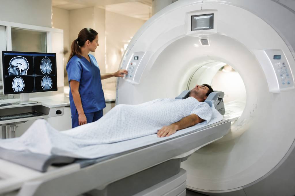
Magnetic resonance imaging (MRI) uses strong magnetic fields and radiofrequency energy to produce detailed images, particularly of soft tissues. Clinicians may use it to investigate some muscle, ligament, spinal or brain conditions when the clinical question calls for that level of detail.
MRI does not use ionising radiation, but “no X-rays” does not mean “no safety considerations”. The magnetic field is powerful and always present, including when a scan is not actively running. Ferromagnetic objects can become dangerous projectiles, while some implants, medical devices and metal fragments may be affected by the field or may make scanning unsuitable without careful checks.
People attending an MRI appointment are normally asked about implants, previous surgery, metal exposure and other relevant factors. A worker who may have metal fragments in the eye from grinding or machining should tell the clinical team before entering the MRI area. Employers can help by giving the worker clear information about past exposure, but the imaging service must carry out its own screening.
Noise, confined spaces and the need to remain still can also affect the experience. Tell the clinical team about concerns in advance; they can explain the process and discuss suitable support. Never enter a controlled MRI area with tools, phones, keys or other items unless the MRI team has confirmed they are safe.
Nuclear medicine and PET: imaging function as well as structure
Nuclear medicine examinations use a small amount of radioactive material, known as a radiopharmaceutical, to provide information about how parts of the body are functioning. Positron emission tomography (PET) is one type of nuclear imaging, often combined with CT to show both function and anatomy. These techniques are used for specific clinical questions and are not routine tests for every work-related symptom.
Because radioactive substances are involved, healthcare providers apply specific procedures to manage exposure and protect patients, staff and the public. Patients receive instructions tailored to the examination. If a worker has questions about returning to work after a scan, particularly where their role involves close contact with others or specific radiation-sensitive duties, they should ask the nuclear medicine team for personalised advice rather than relying on general rules.
Thermal imaging and workplace inspections
Not all workplace imaging is medical. Thermal cameras detect infrared energy and display temperature patterns. Facilities teams may use them to identify overheating electrical components, insulation gaps, moisture patterns or hot spots in machinery. A thermal image can help direct an inspection, but it does not prove the cause of a problem on its own.
For example, a warm patch on a switchboard may signal a loose connection, a heavy load or another issue requiring investigation. A competent person should interpret the result and decide what checks or repairs are needed. Thermal cameras do not see through walls, and readings can be affected by surface materials, reflections, distance and environmental conditions. Treat the image as useful evidence, not a magical X-ray vision upgrade.
Other workplace imaging tools include borescopes for inspecting enclosed machinery and cameras used in remote or robotic inspections. These can reduce the need for workers to enter confined or hazardous spaces, but they do not remove the need for a suitable risk assessment, competent operators and safe equipment.
Choosing the right modality: start with the question
Different modalities answer different questions. A clinician deciding how to investigate an injury considers symptoms, examination findings, medical history and the result needed. In workplace inspections, the responsible specialist considers what hazard is being assessed and whether the imaging tool is suitable for the conditions.
Before an examination or inspection, it can help to ask:
- What specific question is the image intended to answer?
- Will the result change treatment, work restrictions, maintenance or another decision?
- Is there a suitable alternative, and what are its advantages and limitations?
- What preparation, screening or access controls are required?
- Who is qualified to operate and interpret the equipment?
These questions are not a challenge to professional expertise. They are a way to understand the purpose of the test and help ensure that relevant information is not missed.
Practical safety steps for employers
When a worker needs medical imaging, employers should focus on timely care, accurate information and a safe return-to-work plan. They should not pressure employees to undergo or refuse a scan, and should respect confidentiality. Medical information should be handled appropriately, with only necessary details shared with those responsible for supporting the worker.
Useful actions include:
- Record clear facts about the incident, symptoms reported and relevant work tasks, without trying to make a diagnosis.
- Tell the healthcare provider about relevant hazards or exposures, such as a fall, impact, repetitive task or possible metal fragments.
- Arrange emergency assistance where the circumstances or symptoms require it; do not wait for an imaging appointment.
- Follow clinical advice on temporary duties and restrictions, using occupational health support where appropriate.
- For workplace inspection equipment, check competence, maintenance, access controls and task-specific risk assessments before use.
A useful example: a maintenance technician reports shoulder pain after repeated overhead work. An image may or may not be clinically indicated. Regardless, the employer can review task duration, reach, load, equipment and work-rest arrangements. Imaging investigates a person; risk assessment examines the work. Often, addressing both gives the most useful outcome.
Make the image part of a bigger safety picture
Imaging can be powerful, but it is only one part of good occupational health and safety practice. A scan does not replace a conversation with the worker, a clinical examination, exposure records or a review of the task that may have contributed to the problem. Nor does a thermal image replace competent inspection and maintenance.
Choose the tool that answers the question, use it under the right safety controls and make sure the result leads to an appropriate next step. That is how imaging becomes more than a striking picture: it becomes useful evidence for better care, safer work and sounder decisions.