


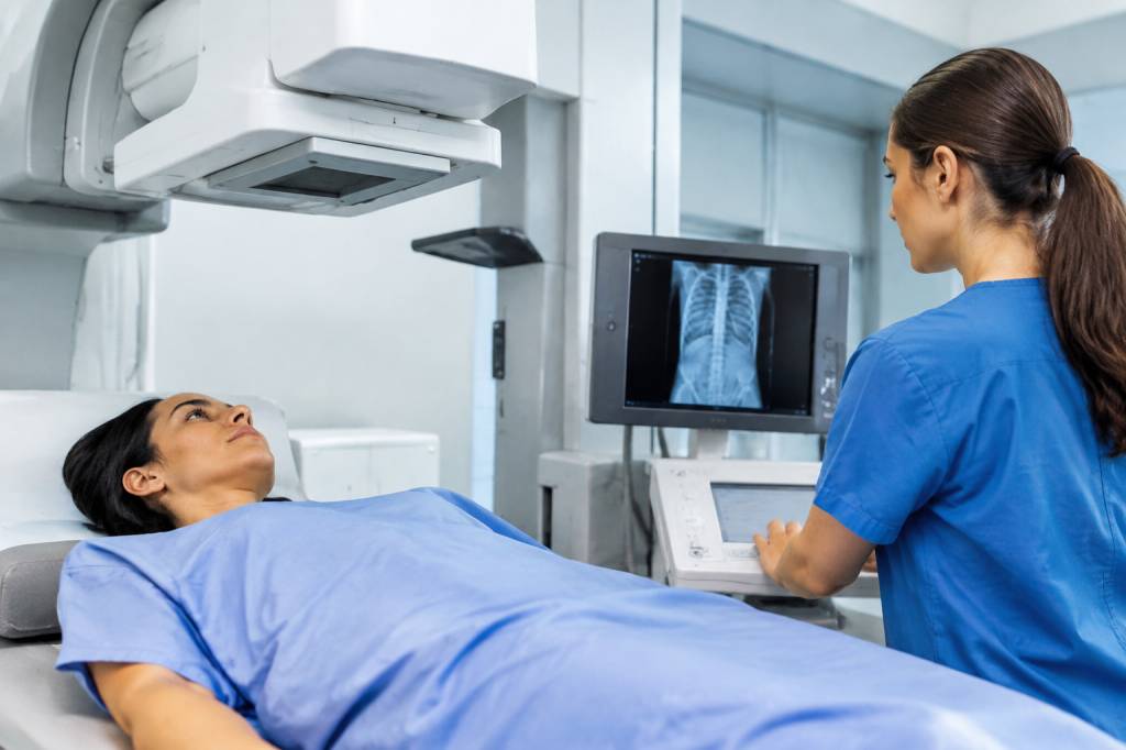
A diagnostic image can look effortless: a patient arrives, the scan takes a few minutes, and a clear picture appears on screen. Behind that picture is a series of careful decisions that protect the patient as well as support an accurate diagnosis. Radiographers are central to those decisions. Their work involves much more than operating imaging equipment; it combines clinical judgement, communication, technical skill and a constant focus on safety.
Whether they are taking an X-ray, performing a CT scan, supporting an MRI examination or working in another imaging service, radiographers help ensure that the right examination is carried out in the right way for the right person. In a busy department, that means managing risk without making patients feel like they have become a checklist with a wristband.
What does a radiographer do?
Radiographers are registered healthcare professionals who produce diagnostic images and, depending on their role and training, may also take on additional clinical responsibilities. They work with patients, radiologists, referring clinicians, nurses, physicists and other colleagues to make imaging safe and useful.
Their responsibilities can include checking a referral, confirming a patient’s identity, explaining an examination, positioning the patient, selecting appropriate imaging settings and checking that the resulting images are suitable for clinical interpretation. They also monitor the patient during the procedure and respond if a concern arises.
The precise scope of practice varies. In the UK, the Ionising Radiation (Medical Exposure) Regulations 2017, commonly known as IR(ME)R, define roles such as referrer, practitioner and operator. A radiographer may act in one or more of these roles when appropriately entitled by their employer. The key point is that responsibilities must be clear, staff must be trained for the tasks they perform, and the patient’s exposure must be properly managed.
Making sure the examination is justified
Medical imaging can provide vital information, but an examination should only go ahead when its expected benefit outweighs the potential risk. This is known as justification. For examinations involving ionising radiation, such as conventional X-rays and CT, the decision must be made by a practitioner who is entitled to do so under local procedures.
Radiographers contribute by checking that the request contains enough information, confirming that the examination matches the clinical question and raising concerns when something does not add up. For example, a referral may request an X-ray of the left wrist while the notes describe an injury to the right. A quick clarification can prevent the wrong examination and the need for a repeat.
If the request is unclear or appears inappropriate, the safest response is not to guess. The radiographer can contact the referrer or an appropriate practitioner to discuss the request. This is a practical example of safety culture: speaking up early, before a small discrepancy becomes a bigger problem.
Identifying the patient and the correct examination
Before imaging begins, the radiographer needs to establish that the patient is who they say they are and that the planned examination is correct. In practice, this usually means asking the patient to state their details and checking them against the referral and local identification procedures. Asking, “Are you Alex Smith?” may prompt a polite nod, but it is less reliable than inviting the patient to say their name and date of birth.
Where relevant, the team also checks the body part and side to be examined. This matters particularly for limbs, where right and left can be easy to confuse in documentation, conversation or a hurried handover. Clear labelling, careful positioning and the use of local checks all help reduce the chance of a wrong-site image.
Patients may be unable to confirm their identity themselves, for example because they are very young, distressed, unconscious or living with communication difficulties. In those situations, staff use the approved alternative identification process, involving records, carers or colleagues as appropriate. The aim is reliable identification, not a one-size-fits-all script.
Explaining the procedure and supporting consent
Safety includes helping patients understand what is going to happen. A concise explanation can reduce anxiety, improve cooperation and make it easier to obtain a useful image. The radiographer should describe the relevant steps in plain language, including what the patient may need to do, how long the examination is likely to take and whether it involves an injection, close positioning or holding still.
Consent is an ongoing conversation, not simply a form signed at the start. Patients should have the opportunity to ask questions and raise concerns. They can also withdraw their consent. If a patient appears uncertain or distressed, pausing to check understanding is usually more useful than trying to power through on the theory that the scanner is already warmed up.
Communication may need adapting for children, people who do not speak English confidently, patients with sensory impairments and those with learning disabilities or cognitive difficulties. An interpreter, accessible information, a support person or extra time may help. Staff should also consider privacy and dignity, especially when a patient needs to change clothing or be positioned in a vulnerable way.
Managing radiation exposure responsibly
For imaging that uses ionising radiation, patient safety depends on using the examination only when justified and keeping exposure as low as reasonably practicable while still producing images that answer the clinical question. This is often described as optimisation. It does not mean choosing the lowest possible exposure regardless of image quality; an image that cannot support diagnosis may lead to a repeat examination and further exposure.
Radiographers use their knowledge of the patient, the equipment and the examination to select appropriate techniques. Factors such as the body area, patient size, positioning and clinical purpose all matter. Correct positioning is particularly valuable: it can help avoid unnecessary images and reduce the likelihood of repeating an examination because anatomy was missed or obscured.
For a patient who may be pregnant, local procedures guide how the team asks and assesses relevant questions. The approach depends on the examination and clinical circumstances. Staff should handle these conversations sensitively and avoid assumptions. If there is uncertainty, the examination can be discussed with an entitled practitioner before proceeding.
Radiation protection also involves staff and the wider public. Equipment checks, controlled areas, appropriate shielding practices where indicated, and adherence to local rules all contribute. The objective is not to make imaging frightening; it is to use a powerful diagnostic tool thoughtfully.
MRI safety: the checklist really matters
MRI does not use ionising radiation, but it is not risk-free. Its strong magnetic field can attract certain objects with considerable force, and some implants or devices may be affected by the examination. That is why MRI screening is a clinical safety step, not administrative paperwork designed to test a patient’s patience.
Radiographers check for relevant implants, previous operations, metal fragments, devices and other circumstances that may affect whether or how an MRI can be performed. Items such as phones, keys, tools and some forms of equipment must be kept out of the controlled MRI area. Patients should be encouraged to mention anything they are unsure about rather than deciding for themselves that it is probably fine.
When a patient has an implant or device, staff follow the relevant safety information and local procedures. They may need to confirm the exact device, assess its conditions for scanning or seek specialist advice. A screening concern should be resolved before entering the scanner room, where the magnet does not accept “I thought it would be all right” as a safety strategy.
Contrast agents and patient monitoring
Some imaging examinations use contrast agents to make particular structures or abnormalities easier to see. Before administration, the team considers relevant risks and follows local protocols, which may include checking a patient’s history, medication, allergies or kidney function according to the agent and clinical circumstances.
Radiographers and other appropriately trained staff explain what the patient may experience and monitor them in line with the procedure. Some people notice a brief warm sensation or an unusual taste after an injection; others may have no noticeable effect. If a patient reports pain, swelling, breathing difficulty or another unexpected symptom, staff need to respond promptly and follow the department’s escalation arrangements.
For example, if a patient says that their arm feels increasingly tight during an injection, the team should not dismiss it as ordinary nerves. The injection may need to be stopped and the patient assessed. Clear instructions before the scan—such as telling patients to report pain or discomfort straight away—make this kind of response easier.
Infection prevention and equipment safety
Imaging departments see people with a wide range of health needs, so infection prevention must be part of routine practice. Radiographers follow hand hygiene requirements, clean equipment and contact surfaces according to local policy, and use personal protective equipment when indicated. A patient table may look spotless, but appearances are not a substitute for a reliable cleaning routine.
Equipment safety matters too. Imaging systems require appropriate maintenance and quality assurance, while staff need to recognise and report faults. If a detector, table or positioning aid is not working as expected, improvising around the problem can create avoidable risk. Reporting the issue helps protect patients and colleagues, and allows the equipment to be assessed before it is used again.
Keeping patients safe during positioning and transfers
Positioning a patient can involve moving them onto a table, supporting an injured limb or helping someone remain still for several minutes. Radiographers assess what the patient can manage and adapt the approach where needed. Pain, reduced mobility, dizziness, frailty and the risk of falling should all be considered.
For a patient who has difficulty standing, for instance, a chair, transfer aid or additional staff support may be safer than a quick lift. The right choice depends on the patient and local moving-and-handling guidance. Radiographers should also explain before touching or repositioning someone, check comfort where possible and protect lines, drains or other medical devices.
Small adjustments can make a real difference. A cushion or support may reduce discomfort, while clear instructions can help a patient hold still without feeling rushed. Comfort is not a luxury: a more comfortable patient is often better able to cooperate, which can improve both the experience and the image.
Communication, teamwork and learning from incidents
Safe imaging relies on accurate information moving between people. Radiographers may need to share relevant concerns with a referrer, radiologist, nurse or other member of the care team. They also need to document examinations and follow local processes for unexpected findings, delays, technical problems or incidents.
When something goes wrong or nearly goes wrong, reporting it supports learning. An incorrect referral caught before imaging, a near miss involving an MRI safety check or a repeated image caused by a positioning issue can all reveal where a process could be stronger. The purpose of a constructive review is to understand what contributed—not simply to find someone to blame.
Consider a department where patients are regularly arriving without clear information about mobility needs. The solution may involve improving referral prompts or handover arrangements, rather than expecting each radiographer to discover the problem at the scanner door. Good systems make the safe action easier to take consistently.
What patients can do to help
Patients are partners in their own safety. They can help by bringing relevant information, answering screening questions as accurately as possible and speaking up if something seems wrong or uncomfortable. It is always reasonable to ask why an examination is needed, what it involves or whether there are alternatives.
- Confirm your identity and the examination details when asked.
- Tell the team about implants, devices, previous reactions or relevant medical information.
- For MRI, mention any metal or device you are unsure about before entering the scanning area.
- Report pain, discomfort, anxiety or unexpected symptoms during the examination.
- Ask for an explanation if you do not understand an instruction.
Radiographers bring together technical expertise and human care at a moment when patients may feel worried, unwell or unsure. By checking details, applying safety standards, adapting to individual needs and communicating clearly, they help make diagnostic imaging both useful and responsible. The best safety measures are often the ones patients barely notice—because someone has already thought carefully about what could go wrong and how to prevent it.